
Presentation
The patient presented with left anterolateral neck swelling for 3 years and recently developed a headache.
Patient Data


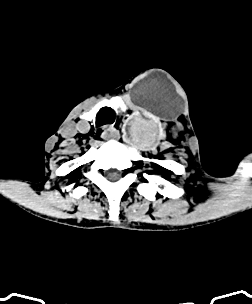
The left common carotid artery (CCA) has tubular dilatation throughout its course, from its origin in the aortic arch to the carotid bifurcation, and is filled with heterogeneous, non-enhancing intraluminal content, inferiorly hyperdense and superiorly fluid density lower signal material. The hyperdense thrombus content fills the proximal and mid-CCA. The left vertebral artery is pushed posteriolaterally without haemodynamically significant change.
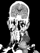
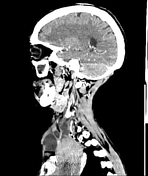
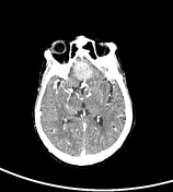
Additionally, there is a hyperdense lesion at the planum sphenoidale with perilesional hypoattenuation and post-contrast homogenous enhancement, almost certainly a planum sphenoidale meningioma.
Conclusion
Total occlusion of the left common carotid artery with pseudoaneurysm from its origin in the aortic arch to the carotid bulb. Retrograde flow contrast fills both the external carotid artery (ECA) and the internal carotid artery (ICA).
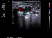
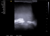

Grey-scale images of left CCA show the lumen to be filled with echogenic thrombus and sluggish turbulence between the clotted and non-clotted blood component within the dilated lumen at the level of mid-CCA. Both ICA and ECA calibre appear normal and have no internal content.
On CCA, colour imaging does not display flow rather artifact colour jet is seen between the thrombus-filled proximal and the non-clotted distal part due to sluggish movement. The internal carotid artery and external carotid artery are both shown as patent on a colour Doppler picture of the left carotid bifurcation. The flow in the ICA is still anterograde, while the flow in the ECA is reversed, according to colour Doppler pictures with spectral Doppler tracings.
Case Discussion
Both ICA and ECA have patent approval, confirmed by contrast filling from reverse flow either through ICA or ECA. Mostly, the ECA will reverse and feed the ICA with blood. There is also reverse flow from ICA that will fill the ECA.
Thanks to its huge pool of possible collaterals, both ECA and ICA reverse flow can replace the totally occluded CCA supply. This fascinating phenomenon causes one vessel to appear on a colour Doppler examination as red and the other as blue.


 Unable to process the form. Check for errors and try again.