
Presentation
Headache, fever, nasal congestion, cough, dyspnoea, pleuritic pain, myalgia, loss of smell, and taste, with the onset of symptoms 11 days ago.
Patient Data


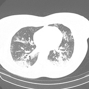
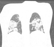
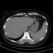
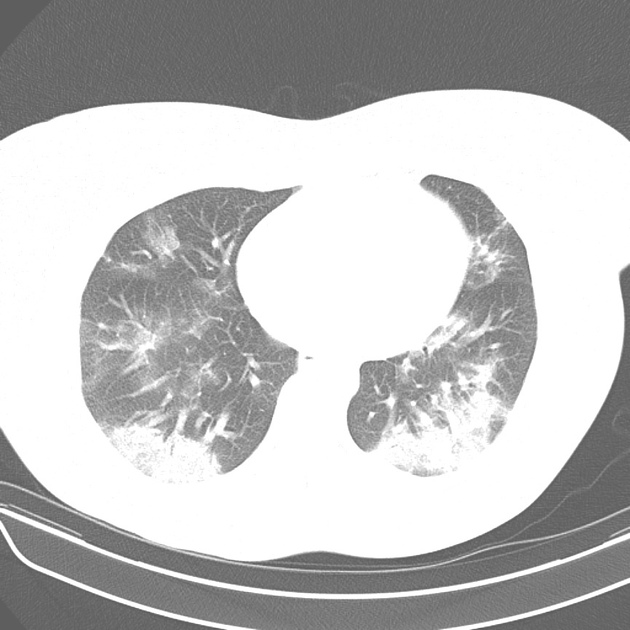
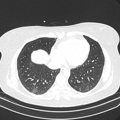
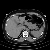
CT shows bilateral, multi-lobar areas of mixed airspace consolidations with ground-glass opacities in both lungs, some of them with reticular and interlobular septal thickening featuring the crazy-paving pattern. The lesions are mostly in the peripheral parts of the lungs. Small bilateral pleural effusion
This patient tested positive for SARS-CoV-2, and the CT findings are consistent for peak stages (stages 3) COVID-19 pneumonia.
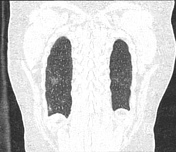

CT reveals regression of the pleural effusion and of the lung abnormalities related to pneumonia COVID-19, leaving rare and tenuous bilateral residual ground-glass focal areas, usually found at the late/absorption stage (stage 4).
Case Discussion
Covid-19 is a disease caused by SARS-CoV-2 1-6. Chest CT can help guide the clinical decision and monitor disease progression 1-6. In stage 3 (peak stage, 9-13 days from onset of the symptoms), consolidations become prevalent concerning ground-glass opacities (GGOs) 1-6. In stage 4 (absorption/late stage, After 14 days), the consolidations are gradually absorbed and replaced by subpleural parenchymal bands and residual faint GGOs, which can persist as long as one month and beyond 1-6.
This case illustrates the temporal CT changes of peak stage (stages 3), improving at the late/absorption stage (stage 4) of COVID-19 pneumonia.


 Unable to process the form. Check for errors and try again.