
Presentation
Out of hospital arrest. For investigation.
Patient Data


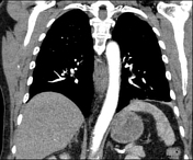
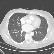
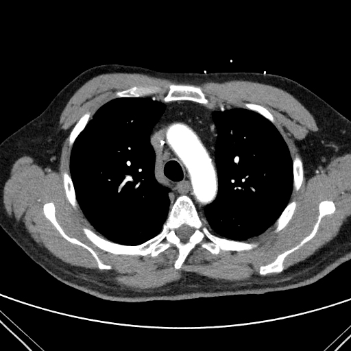
There is a large parenchymal mass in the posterobasal and lateral basal segments of the left lower lobe. The mass has a spiculated border, with a broad pleural abutment. Local peribronchial thickening. Sub-segmental atelectasis medial to the dominant mass.
Small region of patchy mixed ground glass consolidation/collapse the inferior lingula.
Small left pleural effusion.
Rounded but sub-centimeter lymph nodes in the left hilar and bilateral lower paratracheal stations.
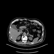
No aggressive skeletal lesions or abnormalities in the upper abdomen.
Bilateral non-displaced anterior rib fractures.
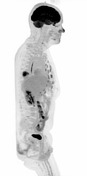
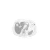
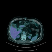

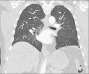
The basal left lower lobe pulmonary mass moderate to intensely FDG avid, with central photopaenia consistent with necrosis. No FDG avid lymphadenopathy or evidence of distant metastatic disease.
Small focus of uptake in the left neck over the sternocleidomastoid muscle, most likely inflammatory. Linear uptake in the distal esophagus is consistent with esophagitis.
Focal intense uptake in the mid sigmoid colon (left pelvis) concerning for an underlying polyp.
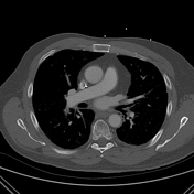
Focal uptake in multiple ribs anteriorly, corresponding to non-displaced fractures on low-dose CT. The pattern is symmetrical and consistent with post-CPR injuries. Focal uptake in the spine centered on an anterolateral osteophyte, reflecting active arthritis.
Case Discussion
Primary malignancies can be diagnosed unexpectedly on imaging performed for other reasons, in this case investigation following cardiac arrest.
F18-FDG is most commonly used for assessing malignancy, however is not specific for cancers. Other physiological and pathology processes which undergo glucose metabolism can also produce scan abnormalities, including infection, inflammation and resolving trauma. Due to the nature of this patient's presentation, traumatic injuries were also hypermetabolic on F18-FDG PET-CT when the newly diagnosed lung cancer was staged. The PET-CT was helpful in this case as indeterminate nodes on the CT study were not FDG avid.


 Unable to process the form. Check for errors and try again.