
Presentation
A known case of Crohn's disease presented with acute abdominal pain.
Patient Data
Age: 10 years
Gender: Female
From the case:
Crohn disease - comb sign


Download
Info
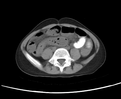
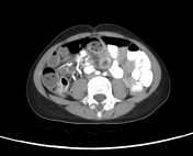
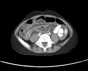
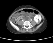
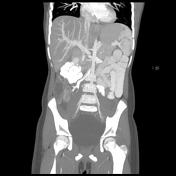
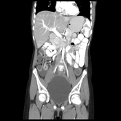
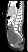
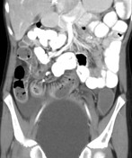
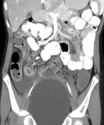
CT study shows the following:
- circumferential mural thickening of multiple loops of the distal ileum with mucosal hyperenhancement. These changes are more prominent at the terminal ileum where a significant mural thickening results in a stenotic lumen with subsequent upstream dilatation results in moderate dilatation of multiple distal ileal loops
- associated congested mesenteric vessels are more prominent at the right iliac fossa resulting in a characteristic "Comb sign". The ileocolic- right colic arterial trunk appears hypertrophied
- blurred mesenteric fat planes, multiple mesenteric lymph nodes, and mild intraperitoneal free fluid collection
- all these changes are radiologically suggestive of an active attack of Crohn's disease

Case Discussion
Here is a case with typical CT findings of Crohn disease including the comb sign.


 Unable to process the form. Check for errors and try again.