
Presentation
Chronic diarrhoea and recurrent abdominal pain.
Patient Data
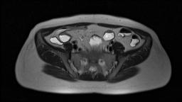

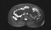
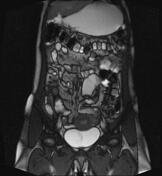
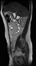

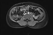
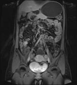
multifocal segmental mural thickening with dominant mesenteric side involvement in the distal & terminal ileum with some skip segments in between
short segment stenosis in the terminal ileum measuring about 3cm in length and extending to the ileocaecal valve with a maximum mural thickness of 9mm & some mural ulcers
intense asymmetric enhancement in the involved ileal segment including the stenotic part accompanied by mesenteric engorgement
prominent pericentric vasculature giving the "comb sign" and prominent fibrofatty infiltration
no evidence of fistula, collection, ascites or obstruction
Case Discussion
MRE imaging findings are in favour of the patient's known Crohn's disease with dominant active patterns in the distal & terminal ileum and short inflammatory strictures at the terminal ileum proximal to the ileocaecal valve.


 Unable to process the form. Check for errors and try again.