
Presentation
Asymptomatic. Patient came for screening and incidental finding of renal ectopia in ultrasonography for which she was planned CT urography.
Patient Data
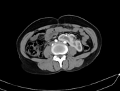

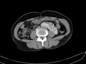
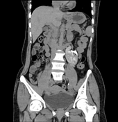
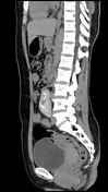

Right-to-left crossed fused renal ectopia is seen, with the upper pole of the right ectopic kidney fusing with the lower pole of the left kidney.
The left kidney is supplied by the left renal artery arising from the aorta, and the right kidney is supplied by the left common iliac artery near its bifurcation from the aorta.
The left renal vein is draining into the IVC, and the right renal vein is draining into the IVC near its bifurcation. The renal pelvis of both kidneys is directed anterolaterally. Both kidneys show prompt contrast enhancement and excretion.
The pelvicalyceal system is not dilated.
No calculus is seen.
A well-defined, non-enhancing fluid attenuation lesion with a thin imperceptible wall measuring approximately 6 mm x 6 mm is seen in the upper pole. No calcification, septations, or enhancing solid components are seen. Both ureters are inserting into their orthotopic position.
Impression:
normal excreting both kidneys
right-to-left crossed fused renal ectopia
left renal cortical cyst (Bosniak I)
Case Discussion
Crossed fused renal ectopia is a rare congenital abnormality of the renal system where one kidney crosses over to the opposite side and fusion of the renal parenchyma takes place. Although left-to-right ectopia is three times more common, here we present a case of right-to-left inferior crossed fused renal ectopia. Patients are usually asymptomatic and are typically diagnosed incidentally. However, there is an increased probability of nephrolithiasis formation in these patients.


 Unable to process the form. Check for errors and try again.