
Presentation
Abdominopelvic pain and microscopic hematuria.
Patient Data
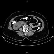

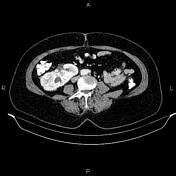
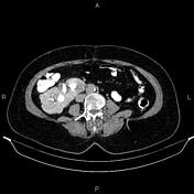
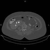
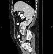
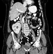
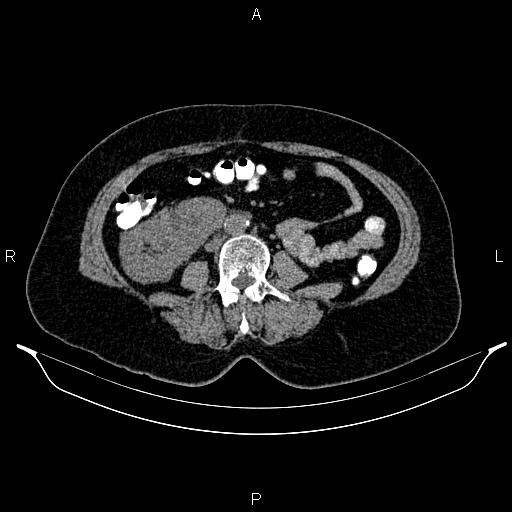
The left kidney has not crossed the mid-line and lies on the right side, inferomedial to the right kidney and fused to the lower pole, inferring crossed fused renal ectopia.
The right renal collecting system notes a 14 mm stone with mean attenuation values of 751HU.
A 28mm calyceal diverticulum is observed in the fused part of the kidneys that contain several hyperdense stones less than 11mm and with mean attenuation values of 7311HU. In addition, a 10mm stone with mean attenuation values of 967HU is evident in the proximal part of the left ureter, which causes mild hydronephrosis.
Grade I spondylolisthesis of L5 on S1 is present.
Case Discussion
Crossed fused renal ectopia with calyceal diverticulum and nephrolithiasis.


 Unable to process the form. Check for errors and try again.