CT angiogram sign - lung adenocarcinoma
Updates to Case Attributes
Updates to Study Attributes
The right lower lobe bronchus is infiltered, and collapse consolidation of the right lower lobe is evident. A large ill-defined infiltrative mass encases the right hilar structures and extends into the adjacent mediastinum. After contrast media administration,vessels appear prominent as they traverse an airless low attenuation portion of the consolidated lung, resembling CT angiogram signs.
A few subcarinal and right paratracheal lymphadenopathy have a short axis diameter of less than 20 mm.
Mild left to right shifting of the heart and mediastinum is also seen.
Image 84 CT (C+ arterial phase) ( create )
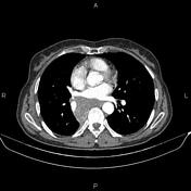
Image 84 CT (C+ arterial phase) ( create )
Updates to Study Attributes
There is a sizeable heterodense mass lesion at the medial margin of the right lung's lower lobe, extending to the mediastinum and subcarinasubcarinal area. It involves the bronchovascular bundle of the lower lobe, leading to extensive peripheral atelectasis and collapse and showing intense FDG uptake (SUVmax=18.6).
The mediastinum has an enlarged lymph node with very intense FDG uptake (SUVmax=22.65) in the right upper paratracheal area. Another enlarged lymph node with similar characteristics is noted in the subcarina.


 Unable to process the form. Check for errors and try again.