
{"current_user":null,"step_through_annotations":true,"access":{"can_edit":false,"can_download":true,"can_toggle_annotations":true,"can_feature":false,"can_examine_pipeline_reports":false,"can_pin":false},"extraPropsURL":"/studies/125210/annotated_viewer_json?c=1660503608\u0026embed_domain=external.radpair.comfavicon.icoradiopaedia-icon-144.png\u0026lang=us"}
Superficial Layer
- Skull base to clavicle
- Encircles entire neck
- Suprahyoid neck: carotid sheath
- Infrahyoid neck: surrounds strap muscles, sternocleidomastoid and trapezius muscles. Carotid sheath.
Download
Info
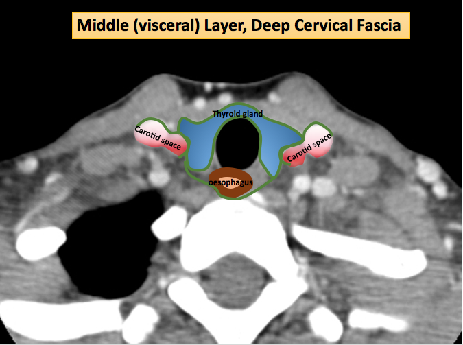
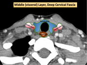
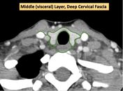
Middle Layer:
- Suprahyoid neck: Buccopharyngeal fascial ; Carotid sheath
- Infrahyoid neck: Visceral fascia
- Encircles "Viscera"
- Posterior margin = anterior wall of retropharyngeal space.

Download
Info
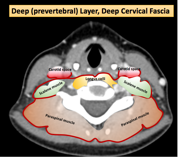
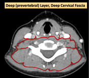
Deep Layer
- Surround perivertebral space; carotid sheath
- Perivertebral space: 1. prevertebral 2. paraspinal
- Encircles prevertebral, scalene, paraspinal muscles
- Lateral wall of retropharyngeal space (Alar fascia)
Case Discussion
Annotated diagrams for outlining the superficial, middle and deep layers of deep cervical fascia.
Each layer contributes to the carotid sheath.


 Unable to process the form. Check for errors and try again.