
Presentation
Seizure
Patient Data
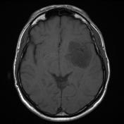
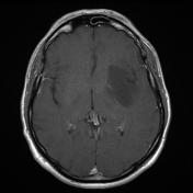
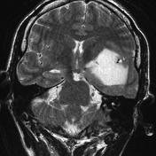
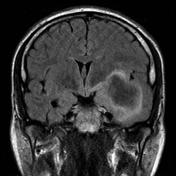
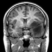
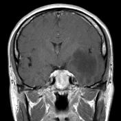
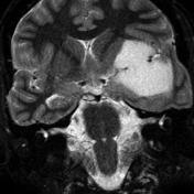
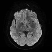

A low T1, high T2 signal mass is present on the left centered on the insular cortex with mild positive mass effect. No enhancement.
Case Discussion
The patient had a craniotomy and resection of the mass.
Histology
Several fragments of white matter are hypercellular and show loss of the normal white matter architecture. Within these fragments, there is a proliferation of mildly atypical astrocytic cells. These have enlarged irregularly shaped hyperchromatic and vesicular nuclei and delicate processes. No mitotic figures are seen and there is no evidence of vascular endothelial cell proliferation and no necrosis. Prominent perineuronal secondary structuring is noted in some areas of cortex.
Final diagnosis: Diffuse astrocytoma (WHO grade II).
Note: IDH mutation status is not provided in this case and according to the current (2016) WHO classification of CNS tumors, this tumor would, therefore, be designated as a diffuse astrocytoma NOS.


 Unable to process the form. Check for errors and try again.