
Presentation
HIV positive patient presented with cerebellar ataxia and signs of raised intracranial pressure.
Patient Data
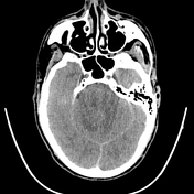

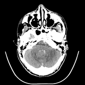

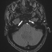
CT scan shows obstructive hydrocphalus secondary to a mass compressing the fourth ventricle. Surrounding vasogenic edema. Mass is isodense to brain parenchyma. Diffuse sulcal and basal cistern effacement consistent with raised intracranial pressure.
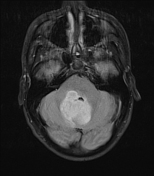
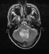
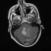
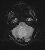
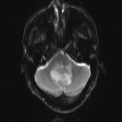
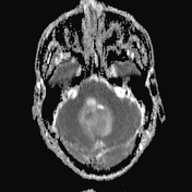
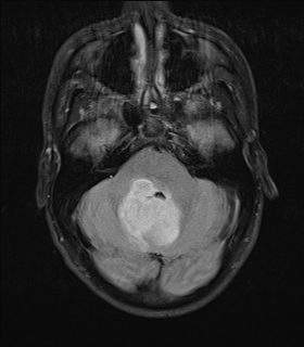
Mostly solid mass involving the cerebellum with some cystic components. Compression of the fourth ventricle with resultant obstructive hydrocaphalus. Sulcal and basal cistern effacement in keeping with raised intracranial pressure. The mass exhibits increased T2 signal, low T1 signal and heterogenous enhancement. No calcifications or hemorrhage. Heterogenous facilitated diffusion of focal solid areas with internal restricted diffusion. Ependymal enhancement involving the fourth ventricle. Normal arteriogram. Biopsy recommended.
Histology
The patient went on to have a brain biopsy.
Gross examination
The specimen consisted of multiple fragments of brown tissue, of which the largest fragment measured 8 mm in greatest dimension. All tissue processed.
Microscopic examination
Microscopic examination of sections of the tissue fragments shows cerebellum with edema and increased cellularity. The lesion is composed predominantly of cells with mild nuclear atypia. Scattered cells with hyperchromatic and bizarre-looking nuclei are also present. No inflammatory cells, necrosis, increased mitotic activity or microvascular proliferation is identified. Special stains for fungi are negative.
In the presence of good controls, immunohistochemical stains are as follows:
- S-100: negative
- GFAP: positive
- Toxoplasma: negative
The morphological features and immunohistochemical profile of the tumor cells are consistent with a diffuse astrocytoma, not otherwise specified, WHO grade 2.
Diagnosis
Diffuse astrocytoma, not otherwise specified, WHO grade 2
Case Discussion
Ventriculoperitoneal shunt was inserted to manage raised intracranial pressure.
Biopsy was performed of the lesion, which showed features of a diffuse astrocytoma, NOS (WHO Grade 2).


 Unable to process the form. Check for errors and try again.