
Presentation
Admitted with reduced consciousness and cognitive function following a road traffic accident 4 days earlier.
Patient Data
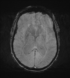
Extensive fine microhemorrhages throughout the cerebral hemispheres and cerebellum with specific conglomeration at the grey-white matter interface, splenium of the corpus callosum and upper brainstem in keeping with a grade III diffuse axonal injury which is important for prognostication.
Case Discussion
MRI brain is useful in the context of traumatic brain injury with reduced consciousness and otherwise normal/subtle CT findings.
The main differentials are diffuse axonal injury and cerebral fat embolism, for which susceptibility-weighted imaging (SWI) is useful. Diffuse axonal injury usually demonstrates microhemorrhages in specific locations such as the grey-white matter interface, corpus callosum and brainstem (regions susceptible to shearing forces due to densely packed fibers). Cerebral fat embolism has a more diffuse distribution of microhemorrhages giving a typical 'starry sky' appearance with suspicions further raised if long bone fractures are present in the body imaging of a polytrauma patient.


 Unable to process the form. Check for errors and try again.