
Presentation
Chronic dysphagia and laryngeal pain.
Patient Data
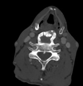

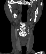
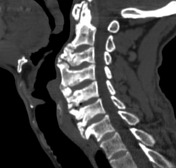
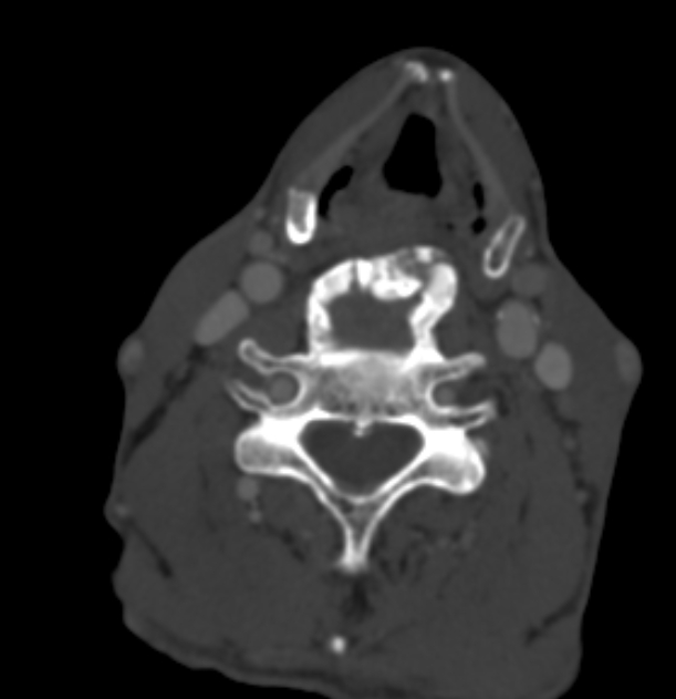
Extensive large anterior osteophytes projecting from CV2 through CV7 vertebrae associated with anterior longitudinal ligament coarse calcifications. This is indenting the cervical esophagus and pushing the larynx anteriorly impressive of diffuse idiopathic skeletal hyperostosis (DISH).
There is also complete ossification of the stylohyoid ligament bilaterally.
3D volume renderig bone window
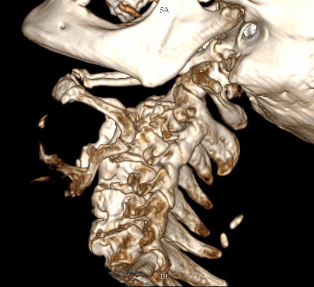
Evident ossification of stylohyoid ligament bilaterally and anterior longitudinal ligament of the cervical spine with large anterior osteophytes.
Case Discussion
CT is a good primary investigation for patients presented with dysphagia, especially solid type, to exclude possible causes of extrinsic compression upon the esophagus. The commonest of which are diffuse idiopathic skeletal hyperostosis (DISH) and eagle syndrome. It's possible to find both phenomena coexist together, although rare 1. Eagle syndrome, if symptomatic, can present also with vascular and neurological compression manifestations.


 Unable to process the form. Check for errors and try again.