
Presentation
The patient complains of chronic neck pain and stiffness, with difficulty moving his head and bending his neck.
Patient Data
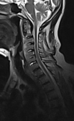

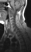

The sagittal MRI of the cervical spine shows prevertebral bone formation and soft tissue ossification, forming a bridge of bone connecting the anterior parts of the cervical vertebrae from C3 downward until T1 vertebra. The disc spaces are more or less preserved.
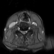
The axial MRI images show that the bone formation is anterior and anterolateral to the vertebral bodies.
There are also degenerative changes of the atlantoaxial joint.
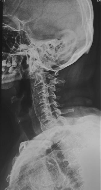
The lateral radiograph of the cervical spine shows a long bone bar and extensive ossification bridging the anterior parts of the vertebral bodies from C2 to T1.
Case Discussion
In this case, the ossification involves six contagious vertebrae (more than three contagious vertebrae) with almost preserved disc spaces are characteristic features of diffuse idiopathic skeletal hyperostosis (DISH). These findings are shown on both the MRI and the lateral cervical spine radiograph.


 Unable to process the form. Check for errors and try again.