
Presentation
Young ballet dancer with severe pain for six months only on the posteromedial aspect of the distal femur. No history of trauma.
Patient Data
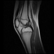


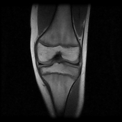


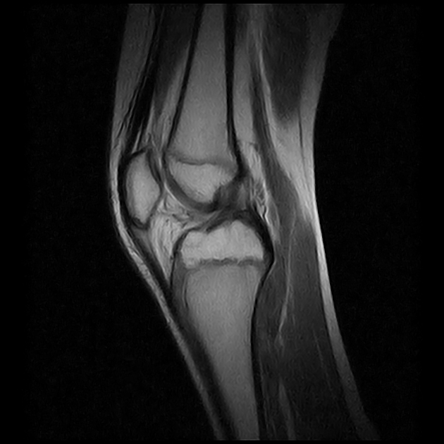
Scalloped lesion with a thin sclerotic rim along the posteromedial distal femoral metaphysis located at the attachment of the medial head of the gastrocnemius muscle. The lesion has low signal on T1 weighted images and intermediate to high signal on T2 and STIR sequences. Enlarged medial gastrocnemius-semimembranosus recess (Baker's cyst). Normal menisci, cruciate ligaments and collaterals. Focal proximal patellar tendon thickening with increased signal intensity as tendinopathy.
A biopsy was performed.
Referto Istologico
Biopsia di lesione litica del femore distale sospetto desmoide corticale. Tessuto fibroblastico con trabecole di tessuto osseo con file di osteoblasti e metaplasia dello stroma fibroblastico. Riassorbimento dell'osso corticale e spongioso da parte degli osteoclasti. Conclusioni: variante di periostite ossificante da proliferazione di tessuto fibroso con neoformazione ossea.
Translation from Italian to English
Histological report
Fibroblastic tissue with trabeculae of bone tissue with osteoblasts and metaplasia of the fibroblastic stroma. Reabsorption of cortical and cancellous bone by osteoclasts. Conclusions: variant of ossifying periostitis from proliferation of fibrous tissue with bone neoformation.
Case Discussion
Distal femoral cortical irregularities (cortical desmoids) are the result of an avulsive injury or stress reaction in the insertion of the medial head of the gastrocnemius muscle or the adductor magnus muscle.
Radiographer: TSRM Luca Piccioni, TSRM Fabio Imola.


 Unable to process the form. Check for errors and try again.