
Presentation
Growth retardation
Patient Data
Age: 12 years
Gender: Female
From the case:
Ectopic neurohypophysis


Show annotations
Download
Info
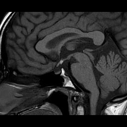
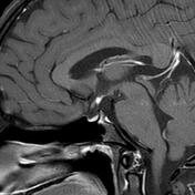
The neurohypophysis shows as bright spot on T1W, located at posterior aspect of the pituitary fossa.
The ectopically located neurohypophysis demonstrates normal enhancement in post contrast study.
Case Discussion
An Ectopic posterior pituitary reflects a disruption of normal embryogenesis of the posterior pituitary and is one of the more common causes of pituitary dwarfism.
MRI findings:
high T1 signal 3-8 mm tissue nodule at the median eminence (floor of the third ventricle)
The exact cause of high T1 signal is uncertain and may relate to neurosecretory granules or high lipid content of the posterior pituitary tissue.


 Unable to process the form. Check for errors and try again.