
Ectopic tooth in maxillary antrum with secondary chronic sinusitis
Presentation
Left mucopurulent nasal discharge with headaches.
Patient Data
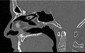

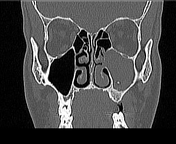
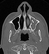

The left maxillary sinus is nearly totally opacified with erosion of its walls and obstruction of the maxillary ostium. A dentiform structure is noted lying within the maxillary antrum.
No bony defect through the floor of the maxillary sinus
Normal pneumatization of the other paranasal sinuses.
Case Discussion
CT features of a chronic maxillary sinusitis secondary to an ectopic tooth in the maxillary antrum.
Ectopic tooth outside the oral cavity is rarely described in the literature, especially within the maxillary sinus. The exact etiology is poorly understood and can be due to defects in embryological development, uneven teeth, genetic factors, trauma, and iatrogenic/idiopathic factors 1,2.
Ectopic teeth are usually asymptomatic, but sometimes may be a source of orofacial pain (impacted teeth), temporomandibular joint pain, or a source of infection such as purulent sinusitis1,2 (in maxillary location, as in this case).


 Unable to process the form. Check for errors and try again.