
Presentation
Pelvic pain with history of previous caesarean section and laproscopic intervention
Patient Data
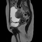

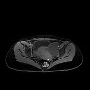
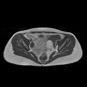
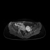

left adnexal lobulated lesion is seen, eliciting a high T1 signal with a low T2 signal (shading sign). It shows no signal drop in the Fat Sat images (left adnexal endometrioma)
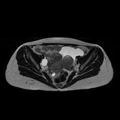
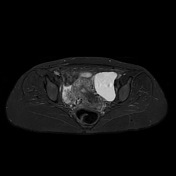
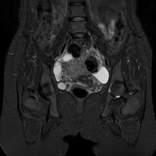
right adnexal lesion elicits a fluid signal of high T2/STIR and low T1 signal.
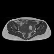
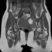
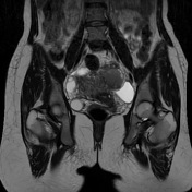
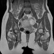
a well-defined, loculated lesion is seen along the medial aspect of both rectus abdominis muscles with haemorrhagic nature (scar endometriomas). A similar lesion is observed at the deep subcutaneous aspect along the anterolateral aspect of the pelvis, abutting the right rectus abdominis muscle at the pelvic region
large left pelvic cystic lesion elicits a fluid-like signal. A smaller similar lesion is seen at the right deep pelvis (peritoneal inclusion cysts)
Case Discussion
the presence of a left adnexal endometrioma in a patient who has had a prior Caesarian section should prompt a search for scar endometrioma
peritoneal inclusion cyst is another complication of previous intervention


 Unable to process the form. Check for errors and try again.