
Presentation
Left parotid region lump for the last few months. Recent onset of local pain.
Patient Data
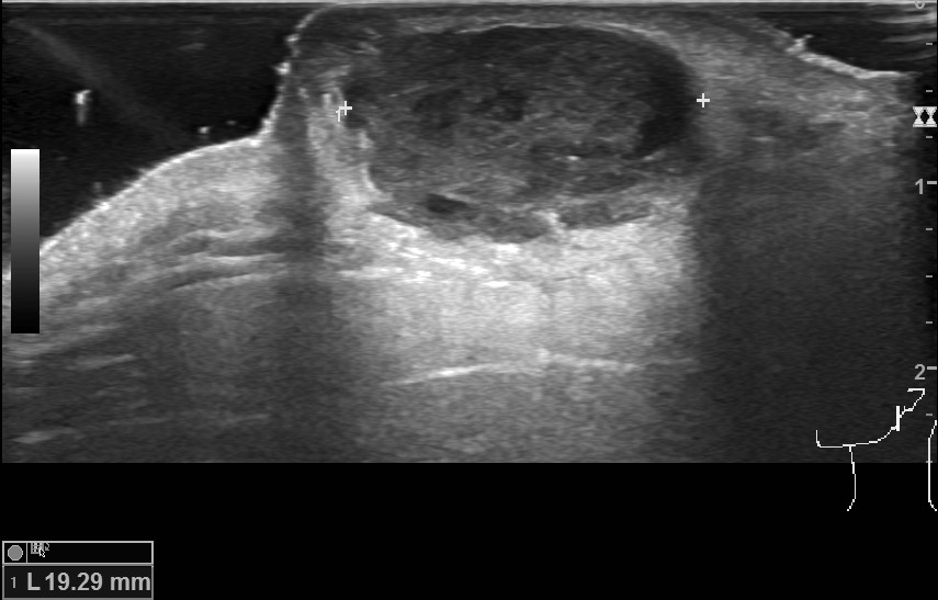
The left parotid region shows an encapsulated lesion superficial to the platysma. It measures about 19 x 18 x 11 mm. It shows a tiny extension to the skin. The lesion reveals a heterogeneous hypoechoic echopattern with tiny cystic space without calcification / internal vascularity. Acoustic enhancement is present. There is a capsule breach on the cranial side of the lesion. The perilesional subcutaneous fat is echogenic with mild hypervascularity.
The left submandibular and superficial lobe of the left parotid glands are normal. There was no cervical lymphadenopathy.
Case Discussion
An elderly male presented with a parotid region lump for the last few months which became painful during the last few days. Ultrasound shows lesion location superficial to platysma which helps to exclude the parotid gland lesion. Ultrasound features favor a leaked epidermal inclusion cyst which was confirmed by the surgery.


 Unable to process the form. Check for errors and try again.