
Presentation
Cerebellar symptoms
Patient Data
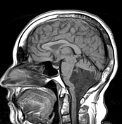

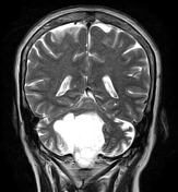
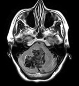
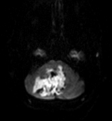
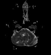
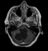
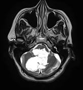

There is an extra-axial mass of irregular lobulated margins centred on the 4th ventricle and extending to the cisterna magna, foramen magnum as well as to the median and lateral apertures. It displays a low signal on T1, heterogeneous/dirty signal on FLAIR, and high signal on T2WI with no enhancement on the postcontrast sequence. It shows a high signal on DWI with low ADC (restricted diffusion).
A mass effect is noted on the brainstem and cerebellar hemispheres with mild cerebellar atrophy.
Case Discussion
Typical MRI features an epidermoid cyst of the posterior cerebral fossa. DWI an important sequence to differentiate an epidermoid cyst from an arachnoid cyst, in which there is no restricted diffusion in arachnoid cyst.
Additional contributor: A. Ramdani, MD


 Unable to process the form. Check for errors and try again.