
Presentation
Galactorrhea and decreased vision.
Patient Data

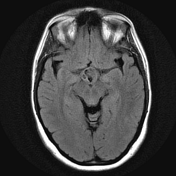
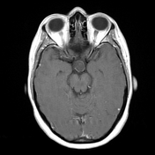
CT is essentially normal, however careful examination reveals expansion of the suprasellar cistern, with CSF density and no calcification.


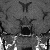
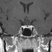
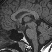
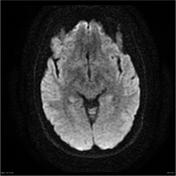
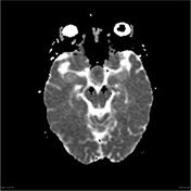

Pre and post contrast scans were performed including dynamic imaging through the pituitary fossa. A complex cystic lesion is seen in the suprasellar region measuring 17 mm transverse, 13mm transverse and 16 mm AP. Internal septation and wall thickening is seen superiorly and as thin rim enhancement post administration of contrast. There is internal restricted diffusion within the lesion. The lesion markedly compresses the optic chiasm which is bowed superiorly around the lesion. There is moderate mass effect on the floor of the third ventricle and hypothalamus. The cavernous sinuses have a normal appearance.
This patient went on to have a craniotomy and excision of the lesion.
Histology
The sections show a cyst, which is lined by keratinizing squamous epithelium with focal parakeratosis. The squamous cells show no evidence of dysplasia or malignancy. No adnexal structures are identified. The stroma is fibrous. The features are those of an epidermoid cyst.
FINAL DIAGNOSIS: epidermoid cyst
Case Discussion
This case illustrates fairly typical appearances of an epidermoid cyst, although thin peripheral enhancement is somewhat atypical.


 Unable to process the form. Check for errors and try again.