
Presentation
Presented with new generalized seizure. Recent period of generally unwell / upper respiratory tract infection symptoms.
Patient Data


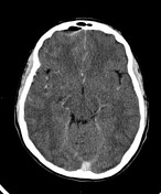
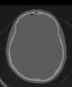
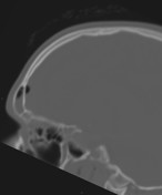
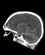
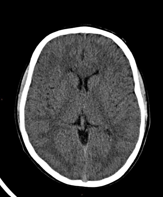
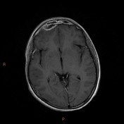
Right frontal peripherally enhancing extradural collection with air, in continuation with frontal sinus opacifcation. The left anterior paranasal and maxillary sinuses are also opacified.
No other intracranial abnormality. No venous sinus thrombosis.
Patient reviewed by ENT and neurosurgery. Given ongoing discharge it was felt surgical drainage would add little benefit and the patient was managed conservatively with broad spectrum antibiotics and antiepileptic medication.
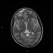
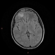
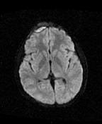
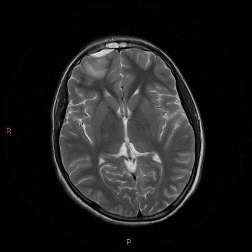
Stable appearance of right frontal extradural abscess. Local dural thickening and enhancement. Restricted diffusion within this lesion. Resolution of intracranial gas. Focally increased T2w / FLAIR signal in the right frontal parenchyma, which may be reactive edema or developing cerebritis. No new collections or areas of signal abnormality.
Case Discussion
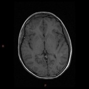
Given good clinical response and stable imaging, the patient was successfully managed conservatively. A three month follow up MRI (not included) showed resolution of the empyema and parenchymal signal change.


 Unable to process the form. Check for errors and try again.