
Presentation
Chronic left foot pain. History of gout.
Patient Data

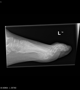
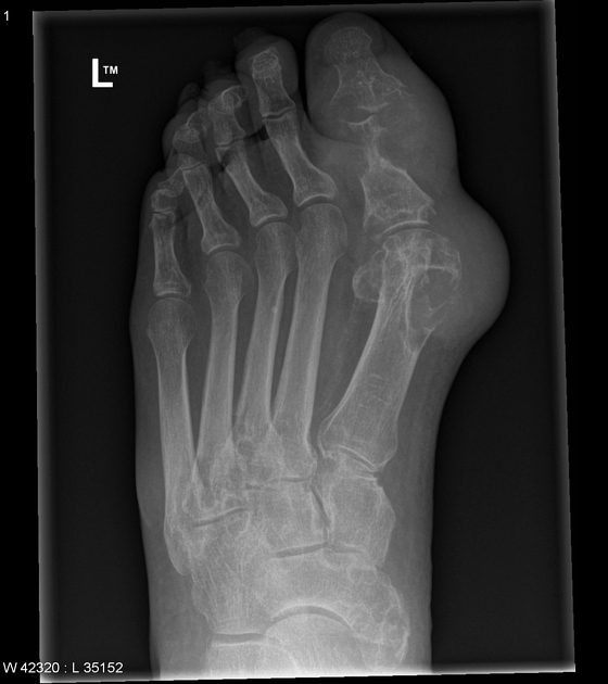
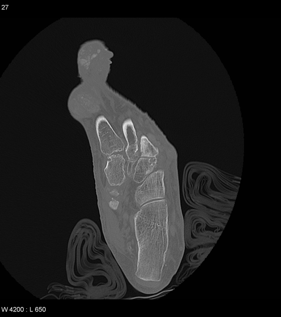
Significant bony erosion of the distal 1st metatarsal, proximal 1st phalanx and base of the distal 1st phalanx with a rat-bitten appearance and overhanging edges. Surrounding soft tissue swelling medial to the erosion with increased density and calcific foci. Erosions involving midfoot and tarsometatarsal joints. Overall, appearances are in keeping with erosive tophaceous gout.
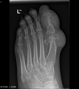
Extensive tophaceous deposits throughout the foot, centred predominantly at the ankle joint, the first metatarsophalangeal joint and the interphalangeal joint of the great toe. There is also a prominent deposit adjacent to the base of the 5th metatarsal.
Besides the rat-bite erosions to the head of the 1st metatarsal and the 1st toe phalanges, coarse erosions scattered throughout the midfoot and tarsometatarsal joints, even involving a type II accessory navicular bone.
Case Discussion
A typical case of erosive tophaceous gout. In particular, a good example of the rat bite erosions that can be seen on the plain radiograph.


 Unable to process the form. Check for errors and try again.