
Presentation
This patient presented to the emergency department with regurgitation, reflux, and several episodes of vomiting after lunch.
Patient Data


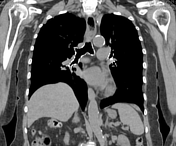
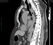
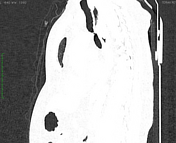
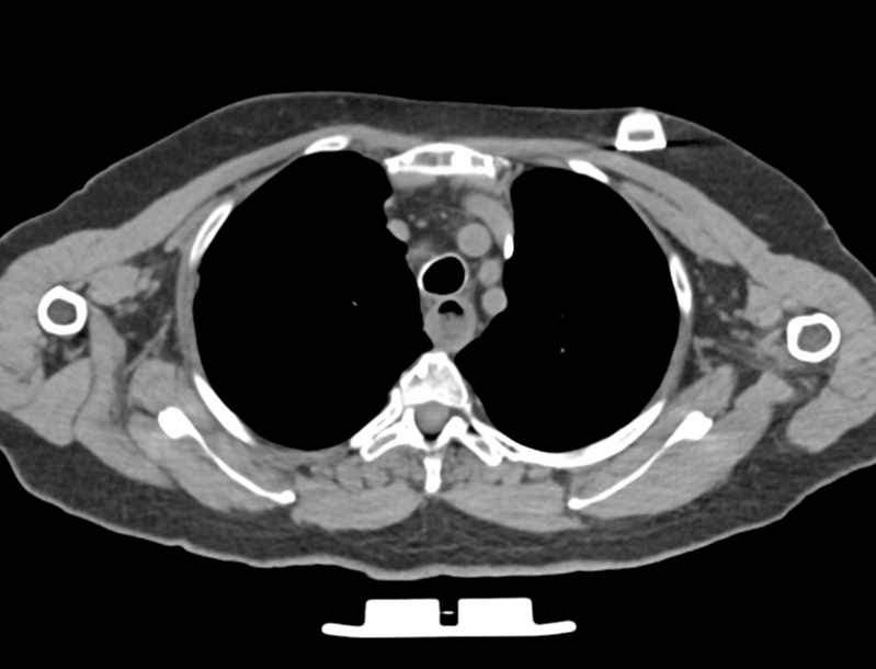
Non-contrast CT scan of the thorax reveals soft tissue attenuation material within the lumen of the thoracic oesophagus at the aortic arch level, a primary area of oesophageal anatomical narrowing. The foreign body measures approximately 1.5 x 2.0 x 3.5 cm. There is focal wall thickening in the oesophagus surrounding the impacted foreign body, indicating inflammation and oedema, and oesophageal dilatation upstream of the foreign body.
CT findings are consistent with an impacted meat bolus in the oesophagus.
Bone lesions are consistent with metastatic bone disease from a previously diagnosed primary breast neoplasm.
The patient underwent an upper gastrointestinal endoscopy.
Gastrointestinal endoscopy
Oesophagus
An impacted foreign body (meat bolus) was observed below the upper oesophageal sphincter. The removal was successful using basket-type forceps. The site of foreign body impaction showed some linear erosions.
Impression: Impacted foreign body (meat bolus) in the oesophagus - successfully removed.
Case Discussion
The oesophagus is a frequent location for foreign body impaction or food obstruction within the gastrointestinal tract 1-6. Although many of these instances resolve spontaneously, some situations require intervention to remove the foreign bodies 1-6. The intervention may involve endoscopic procedures, as in the case of the present patient, who underwent the process and was discharged without complications.


 Unable to process the form. Check for errors and try again.