
Presentation
Decreased apatite, weight loss, and dysphagia for four months.
Patient Data
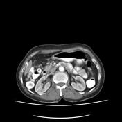

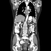
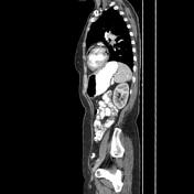
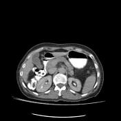
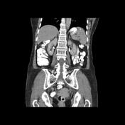
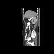

Asymmetrical and circumferential mucosal-wall thickening with thickness up to 12 mm and height up to 48 mm in middle to the distal segment of the thoracic oesophagus is seen and there is not any obvious evidence of aorta or pericardial invasion but a mild impression on the posterior wall of the cardiac left atrium.
Incidental splenic parenchyma and porta hepatis calcifications.
Case Discussion
The case illustrates the contrast-enhanced features of pathology-proved non-metastatic squamous cell carcinoma of the oesophagus. Clinical management is usually based on oesophageal cancer TNM staging and the main treatment is surgical. Early diagnosis is determining outcomes 1.


 Unable to process the form. Check for errors and try again.