
Presentation
Nasal congestion with progressive sinus discomfort/congestion.
Patient Data
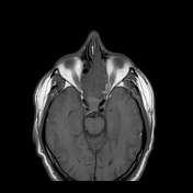

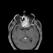
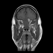
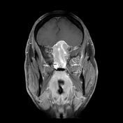
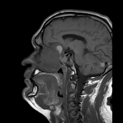
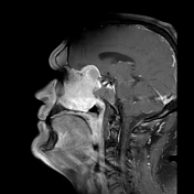
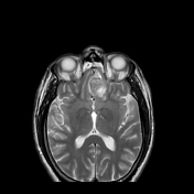
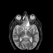
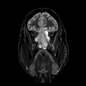

Heterogeneously enhancing soft tissue mass in both sides of the the nasal cavity (left greater than right) and ethmoid air cells with intracranial extension through the cribriform plate and floor of the anterior cranial fossa. Superior displacement of the bilateral frontal lobes (left greater than right). Obstruction of the left maxillary sinus, frontal sinus, bilateral sphenoid sinus, and left posterior ethmoid air cells with T1-hyperintense fluid, suggestive of proteinaceous contents. Medial bowing of the medial wall of the left maxillary sinus and bilateral orbits. On the right, the mass abuts the right internal carotid artery at the level of the clinoid process. On the left, the mass is approximately 1 mm away from the internal carotid artery.
Case Discussion
This is a case of an esthesioneuroblastoma (olfactory neuroblastoma). The patient underwent resection of the mass. Gross pathological analysis revealed a tan-pink rubbery tissue.
Histology
Sections showed a highly atypical cellular proliferation composed of nests or large lobules of round to ovoid tumour cells with enlarged nuclei, frequent vesicular chromatin, distinct nucleoli, and scant cytoplasm. Mitotic figures and foci of tumour necrosis were seen. No neuropil or rosettes architecture were identified. There was perineural and osseous invasion.
The immunohistochemical stains of the tumour cells were strongly positive for synaptophysin , chromogranin , and calretinin, and are negative for AE/AE3 and CAM5.2. The S100 stain highlighted a sustentacular pattern.
Co-authors:
Chris McMahan
Daniel Gewolb, MD


 Unable to process the form. Check for errors and try again.