
Presentation
Patient presented with no neurological signs, recently diagnosed hypercalcaemia. Referral for CT Brain ? basal ganglia calcification.
Patient Data


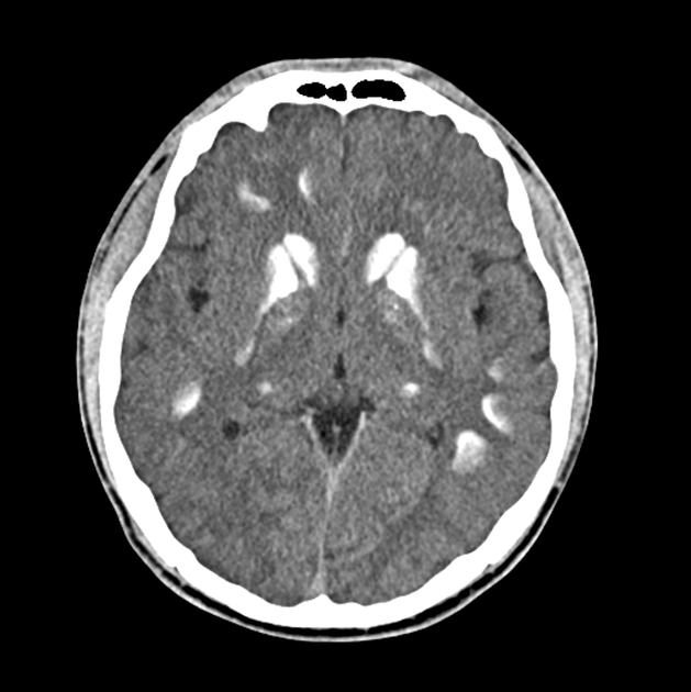
Extensive calcification involving caudate nuclei, putamen and thalamus bilaterally. Calcification involving subcortical grey matter in frontal and parietal lobes. The density measure of calcification was approximately 160 HU.
Case Discussion
Basal ganglia calcification is a rare finding on brain imaging, approximately seen in 1% of all CT brain scans. Rarer still is the finding of this pathology in a person of this age.
There are many differential diagnoses for this appearance, including Fahr syndrome, exposure to toxic chemicals and congenital infection, the latter needs to be strongly considered in this age group.
In this case, the patient was asymptomatic, with hypercalcaemia being the only clinical indicator. In some cases, however, neurological symptoms can be evident and severe, including psychosis, cognitive impairment, dementia.
The DLP for this study was 821 mGy.cm which is below the current ARPANSA reference of 880mGy.cm which is excellent, especially considering patient age. The head is flexed maximally to eliminate direct orbit exposure as much as possible and axial plane reformats performed parallel to the pit fossa-occipital protuberance line (similar to the AC-PC line).


 Unable to process the form. Check for errors and try again.