
Presentation
Progressively worsening hearing loss. Otoscopic examination revealed a mass in the right external auditory canal.
Patient Data
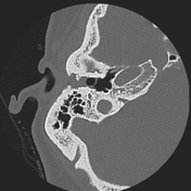

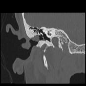
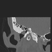
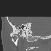

The ear pinna is unremarkable. There is minimal soft tissue thickening along the cartilaginous external auditory canal. However, there is soft tissue density filling the majority of the bony external auditory canal, particularly superiorly. The soft tissue density completely effaces and results in mild erosion of the bony portion of the external auditory canal. There is thickening of the inferior aspect of the tympanic membrane along the pars tensa, as well as minimal retraction of the tympanic membrane.
There is a dysplastic appearance of the lateral semicircular canal and vestibule, noting that the lateral semicircular canal appears small. There is also some patulousness of the vestibule. The cochlea also appears dysplastic, appearing smaller than expected with less than 2 1/2 turns. The anterior scalar septae is absent and the modiolus appears smaller than expected. There is stenosis of the cochlear aperture.
The superior and posterior semicircular canals are relatively normal in appearance. The vestibular aqueduct appears relatively normal in size.
Case Discussion
This is a case of an external auditory canal cholesteatoma, with an incidental note of a cochlear incomplete partition type II. The patient was referred to an otolaryngologist who subsequently performed an excision of the mass. Pathology was consistent with a cholesteatoma. The patient has subsequently followed with otolaryngology without evidence of recurrence.
Co-author:
Luciano Venturino


 Unable to process the form. Check for errors and try again.