
Presentation
The patient presented with a rapidly enlarging left arm mass.
Patient Data


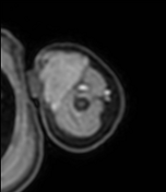
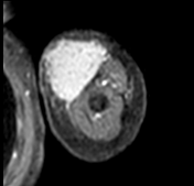
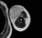
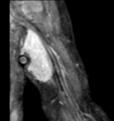
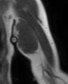
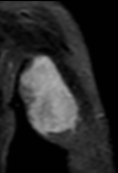
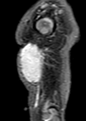
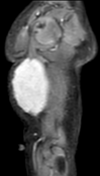
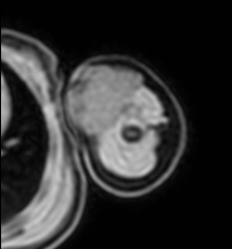
There is a well-defined, homogeneously enhancing soft tissue mass in the subcutaneous fat in the left anteromedial upper arm. The mass abuts the anterior abdominal musculature with no intervening fat plane and has no definite intramuscular extension.
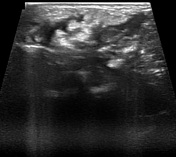
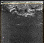

There is a diffusely hypoechoic mass with interspaced hyperechoic and isoechoic foci which appear to represent areas of microcalcifications and interspersed subcutaneous fat. The mass is hypovascular overall, with a vascular appearance similar to adjacent musculature.
Case Discussion
The patient underwent a core needle biopsy.
Histology
Gross: multiple core needle specimens measuring from 1.4 to 2.5 cm in length.
Microscopy: characteristic triphasic growth pattern with bland fibromatosis-like fibroblastic zones, hyalinized zones with crackling artifacts, and small foci of more primitive mesenchyme, showing myxoid stromal change.
Immunohistochemical stains: ALK1 negative, desmin negative, myogenin negative, Factor XIII A negative, EBV negative, CD34 variable staining non-contributory, beta-catenin non-contributory, S100 highlights fat, smooth muscle actin highlights smooth muscle.
Final diagnosis: fibrous hamartoma of infancy.


 Unable to process the form. Check for errors and try again.