
Presentation
Screening US.
Patient Data

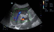
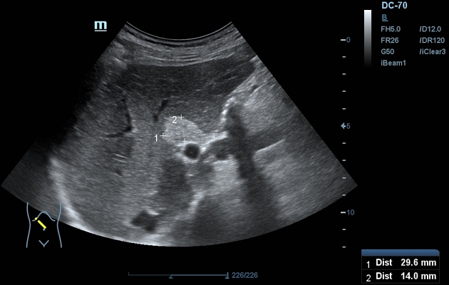
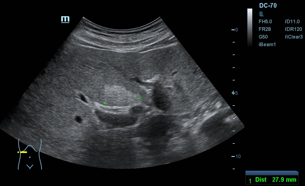
A circumscribed area of increased echogenicity is visible in the liver, immediately anterior to the porta hepatis (segment IV).
The abnormality can be better appreciated on the cine recording.
The abnormality is essentially unchanged compared to the prior exam.
Case Discussion
Segment IV, immediately adjacent to the porta hepatis is the most common location for focal hepatic steatosis.
The ventral aspect of segment IV bordering the falciform ligament is another typical location, and commonly seen with cross-sectional imaging, but only occassionally with US. The likely explanation is that the superficial regions fall into the near field of convex transducers, where contrast resolution is significantly worse.
Focal fatty deposits showing the characteristic appearance and location can be followed with US, whilst for lesions showing atypical location or mass-like appearance, further evaluation with contrast-enhanced US is often warranted. CEUS can rule out malignancy with a specificity on par with cross-sectional imaging, is more cost-effective than MRI, and uses no ionizing radiation.


 Unable to process the form. Check for errors and try again.