Focal nodular hyperplasia
Updates to Study Attributes
The liver exhibits a large iso- to hypodense mass lesion with pronounced contrast enhancement during the arterial phase, observed in the left liver lobe. It manifests a central scar with enhancement on delayed images, suggesting focal nodular hyperplasia.
There is diffuse hepatic steatosis.
Atrophic changes are evident in the left psoas, iliacus, and gluteal muscles, accompanied by diffuse degenerative changes in the left hip joint. These changes are characterizedcharacterised by significant joint space narrowing and subchondral cystic changes. The left acetabulum appears shallow and irregular, indicative of features likely suggestive of neglected developmental dysplasia of the hip (DDH)."
Image CT (non-contrast) ( update )
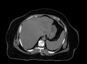
Image CT (C+ arterial phase) ( update )
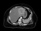
Image CT (C+ arterial phase) ( update )
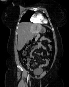
Image CT (C+ portal venous phase) ( update )
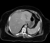
Image 22 CT (C+ arterial phase) ( update )
Image 116 CT (bone window) ( update )
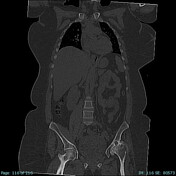
Image 192 CT (C+ arterial phase) ( update )
Image 282 CT (non-contrast) ( create )
Updates to Case Attributes
This incidentally found liver mass with central scar that became indistinguishable (enhanced) on delayed images on a background of a non cirrhotic-cirrhotic liver is most consistent with FNH.
-<p>This incidentally found liver mass with central scar that became indistinguishable (enhanced) on delayed images on a background of a non cirrhotic liver is most consistent with FNH.</p>- +<p>This incidentally found liver mass with central scar that became indistinguishable (enhanced) on delayed images on a background of a non-cirrhotic liver is most consistent with <a href="/articles/focal-nodular-hyperplasia" title="FNH">FNH</a>.</p>
Systems changed:
- Hepatobiliary
- Gastrointestinal