
Presentation
Receptive and expressive dysphasia progressive over 2 years.
Patient Data
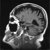

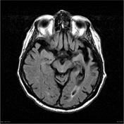
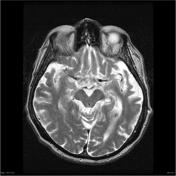
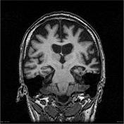

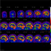
Marked asymmetric atrophy of the anterior left temporal lobe as well as, to a somewhat lesser degree, the left frontal opercular region including the expected location of Broca's area. There is a profound loss of temporal white matter volume with enlargement of the temporal horn. Associated mild increased subcortical FLAIR signal. The rest of the brain, including the hippocampi, demonstrate involutional change, exaggerated for age, but to a much less marked degree.
No intra-cranial mass lesions, acute hemorrhages or collections. No acute or established infarcts. Mild bi-hemispheric chronic white matter ischemic change. Small anterior falcine calcification.
Conclusion:
Findings are in keeping with the language variant of frontotemporal dementia (semantic variant).
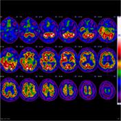
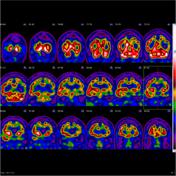
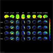
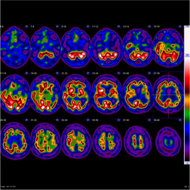
SPECT perfusion studies show severe perfusion reduction in the left temporal lobe with mild perfusion reduction extending into the left frontal and parietal cortex. Right temporal lobe perfusion uninvolved. Posterior cingulate perfusion preserved.
OVERALL IMPRESSION
The severe perfusion reduction involving the left temporal lobe is consistent with language variant of frontotemporal dementia.
Case Discussion
Typical appearances of advanced frontotemporal dementia (semantic dementia)


 Unable to process the form. Check for errors and try again.