
Presentation
Intermittent vomiting with weight loss.
Patient Data


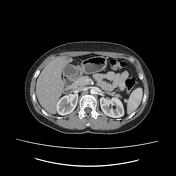
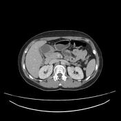
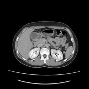
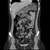
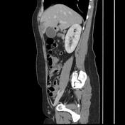
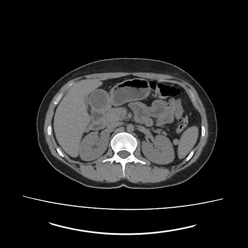
Well-defined bilobulated submucosal cystic mass of the anterior wall of the gastric antrum bulging into the gastric lumen. It shows a homogeneous fluid content with no enhancement on postcontrast images.
Focal fatty infiltration of segment 4 is noted.
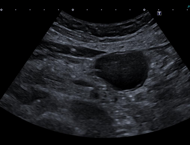
On the ultrasound exam, the cystic lesion shows an echogenic inner layer (mucosal layer) and a hypoechoic outer layer (muscular layer).
Case Discussion
CT features of cystic mass of the gastric wall of submucosal location suggestive of gastric duplication cyst.
The ultrasound exam plays an important role:
to suggest the diagnosis of a gastrointestinal tract duplication cyst by the combination of an echogenic inner mucosal layer and hypoechoic outer muscular layer
to exclude other differential diagnoses such as pancreatic pseudocyst or mucinous cystadenoma or cystic gastrointestinal stromal tumor (GIST).


 Unable to process the form. Check for errors and try again.