
Presentation
Left upper quadrant pain for 3 days, intensified on day of admission. Worsens when eating or moving.
Patient Data


Fat-density structure in the gastrohepatic ligament with a vessel coursing through it and surrounding fat stranding.
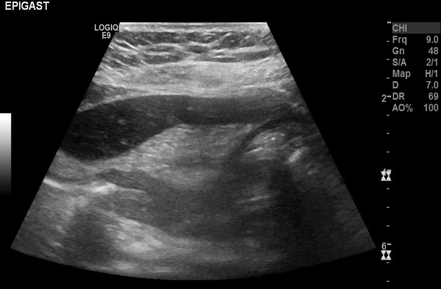
Focal fatty process in the gastrohepatic ligament, containing a hypoechoic focus at its centre, likely representing an infarcted vessel - appearance characteristic of appendagitis and consistent with the finding on the CT study.
Case Discussion
It's not unusual for the radiologist to come up empty-handed when checking an abdominal CT scan for the aetiology of localised pain.
That said, paying special attention to often-neglected anatomical areas (e.g. mesenteries and peritoneal reflections) after the usual suspects have been ruled out can sometimes pay out.
In contradistinction to epiploic appendagitis and omental infarction, perigastric appendagitis 1 is a rare entity. It is also probably underdiagnosed. It should, however, be sought in cases of upper abdominal pain where no other "culprit" has been implicated, especially when blood work comes back normal.


 Unable to process the form. Check for errors and try again.