
Presentation
severe headache
Patient Data
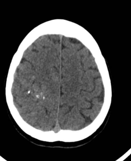
Poorly-defined slightly hyperdense process in upper portion of right cerebral hemisphere, containing serpiginous calcifications concentrated in a small area. Does not exert mass effect. Most likely an AVM.
Tip of basilar artery appears dilated, probably representing an aneurysm.


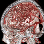
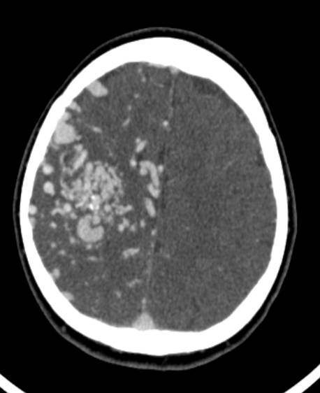
Large arteriovenous malformation (AVM) in right cerebral hemisphere, mostly involving frontal and parietal lobes, with feeding vessels from branches of MCA and ACA and a vein draining into right transverse sinus.
Small basilar tip aneurysm.
Case Discussion
A small cerebral arteriovenous malformation (CAVM) can easily be missed on a non-contrast head CT. In this case, the CAVM was large enough to be detected and the clustered vessel wall calcifications certainly helped narrow down the differential diagnosis.
A small basilar tip aneurysm was also noted; there is an appreciated association between CAVMs and remote aneurysms.
Following detection of the CAVM, a head CT angiogram (CTA) was immediately performed, exquisitely detailing the CAVM.


 Unable to process the form. Check for errors and try again.