
Presentation
Firm, non-tender lump on the left side of the jaw, present for approximately 15 years gradually increasing in size over time. No associated pain or discomfort and no history of trauma to the area. No additional symptoms, such as facial numbness, skin changes overlying the lump, or difficulty in chewing, swallowing, or speaking. No history of fever, weight loss, or recent infections.
Patient Data


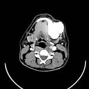
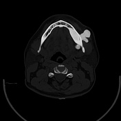
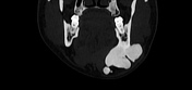
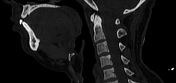
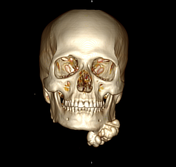
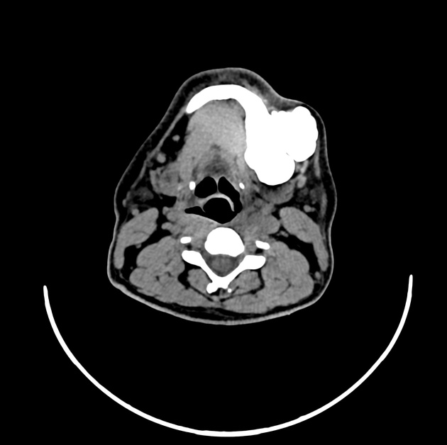
A large, dense, cortical-based bony lesion with a cauliflower-like appearance is present, originating from the body of the mandible on the left side. The lesion measures approximately 50 x 43 x 30 mm and demonstrates a significant mass effect. It displaces the anterior belly of the left digastric muscle medially and pushes the left submandibular gland posteriorly. The mass extends to the subcutaneous plane, causing stretching of the overlying skin. There is no evidence of bone destruction or lytic changes associated with this lesion.
A few small polyps were identified within both maxillary sinuses.
Case Discussion
The lesion's slow, progressive growth over 15 years without associated pain or systemic symptoms, along with imaging findings, strongly favours a benign aetiology. Osteoma is the leading diagnosis due to the dense, well-corticated appearance and the lack of soft tissue invasion, lytic changes, or periosteal reaction. However, further diagnostic steps are warranted to confirm the diagnosis and evaluate any potential impact on surrounding structures, including a biopsy for histopathological examination. Surgical excision may be considered given the mass effect on adjacent soft tissue structures and potential cosmetic or functional impact.
Differential diagnoses:
Take-home message:
Long-standing, painless jaw masses with gradual growth are often benign, but thorough assessment and imaging are essential for accurate diagnosis and management. In cases with significant size or mass effect, as with this patient, surgical excision may be beneficial to relieve pressure on surrounding structures and improve function or aesthetics. Early evaluation and regular follow-up help to prevent complications and ensure optimal patient outcomes.
Case co-authors:
Dr Madan Karmakar
Dr Samir Rana


 Unable to process the form. Check for errors and try again.