
Presentation
Recurrent seizures and headaches.
Patient Data
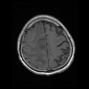

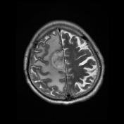
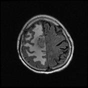
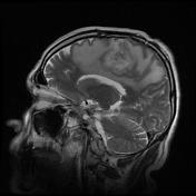
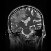


There is a 40*45*50 mm large heterogeneously enhancing tumor involving the right frontal lobe parasagittal region and cingulate gyrus associated with a non-enhancing central fluid signal component suggesting central necrosis.
There is surrounding edema with mass effect on the right lateral ventricle.
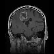
Post-surgical changes of right frontal craniotomy and dural enhancement also are seen.
Case Discussion
This case illustrates the typical appearance of high-grade glioma. The patient went on to have surgery and the diagnosis was confirmed.
Histology
MICROSCOPIC DESCRIPTION: The sections show irregular fragments of crushed brain lesion composed of astrocytic cells with atypical nuclei, many gemistocytes, foci of necrosis and mitotic figures. with regarding the submitted tissue that is crushed and small, making differential histologic diagnosis between anaplastic astrocytoma (gemistocyte rich) and glioblastoma multiform is very difficult.
Final diagnosis: High-grade glioma.


 Unable to process the form. Check for errors and try again.