
Presentation
Preterm baby with low birth weight and birth asphyxia. For routine cranial ultrasound screening.
Patient Data
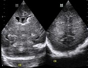

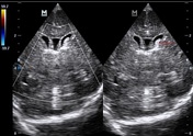

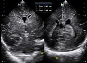
Focal echo-dense, homogeneous material is visualised within the left lateral anterior ventricular horn with the mentioned material showing no vascular signals on colour flow Doppler mapping. There is mild symmetrical fluid-filled dilatation of the lateral ventricles. The brain parenchyma, gyri-sulci pattern, the periventricular white matter and the cerebellum plus the rest of the intra-cranial structures grossly looks normal.
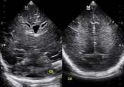
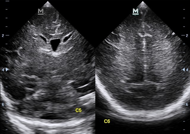
7 days transfontanelle cranial ultrasound follow up shows complete resolution of the previously noted left intraventricular haemorrhage. Bilateral ventriculomegaly is redemonstrated.
Case Discussion
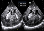
Mild bi-ventriculomegaly accompanied with a focal homogeneously echo-dense focus within the left anterior horn of the lateral ventricle ipsilaterally. The mentioned material occupy an almost <50% of the left anterior horn, lateral ventricular fluid chamber suggesting Grade 2 intraventricular haemorrhage which spontaneously resolved completely on its own without medication 7 days later. Mild reduction in the redemonstrated lateral ventricles' fluid volumes is noted.


 Unable to process the form. Check for errors and try again.