
Presentation
Headache.
Patient Data


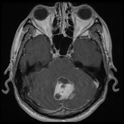
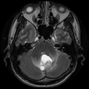
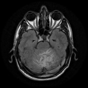
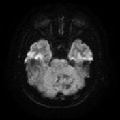
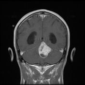
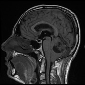

There is a lesion of heterogenous signal intensity arising from the cerebellar vermis extending more to the left side. It is predominantly solid with rim-enhancing cystic component at its inferior aspect. Multiple flow voids at the posterior margin of the lesion suggestive of vascular origin. Severe posterior mass effect at the cerebral aqueduct and fourth ventricle with associated hydrocephalus and transtentorial oedema. There is flattening of the pons and extension of the cerebellar tonsils into the foramen magnum. There is no thickening of enhancement of the dura or diffusion restriction.
Conclusion: demographics and appearance of the mass, particularly the prominent flow-voids, suggests a haemangioblastoma as the most likely diagnosis.
Case Discussion
The patient underwent posterior fossa craniotomy with excision of the lesion.
Histology
MICROSCOPIC DESCRIPTION Sections of the cerebellum show a moderately cellular tumour with scattered stromal cells in a background of many blood vessels of varying sizes. The tumour cells form aggregates. They have enlarged nuclei with hyperchromasia and foamy vacuolated cytoplasm. Mitoses are inconspicuous. There is no necrosis. The tumour cells are focally inhibin positive. CAM5.2 is negative.
FINAL DIAGNOSIS: haemangioblastoma (WHO Grade I)


 Unable to process the form. Check for errors and try again.