
Presentation
The patient presents with a painful left calf mass.
Patient Data


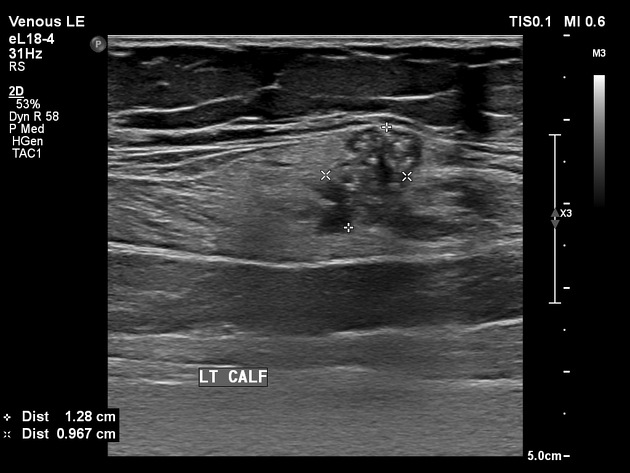
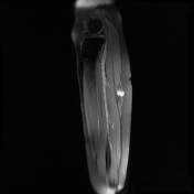
Ultrasound demonstrates a small, irregular, heterogeneous, mixed echogenic mass within the posterosuperior lateral gastrocnemius muscle. Punctate calcifications are present with posterior acoustic shadowing. There is internal vascularity.

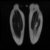

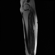
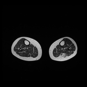
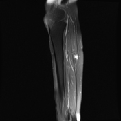

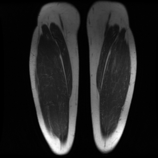
There is a solitary, intramuscular, soft tissue mass within the posterosuperior lateral gastrocnemius muscle measuring 46.9 x 14.2 x 15.9 mm. There is irregular adipose tissue with an eccentric central, apparent cystic component measuring 13.7 x 12.7 x 16.4 mm, with low T1 and high T2 and high Stir signal intensity.
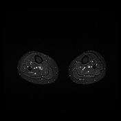
There is a homogenous enhancement of the central component post-contrast administration.
The irregular circumferential adipose components remain non-enhancing.
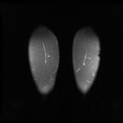
The sonographically identified punctate calcifications are poorly appreciated.
No perilesional oedema or subcutaneous infiltration.
There is no bone infiltration, and no other lesions are identified.
Case Discussion
The MRI features appear typical for an intramuscular myxoma, with an irregular perilesional rim of adipose tissue and eccentric avidly enhancing cystic component. Complete excision histology confirmed the lesion to be a haemangioma (cavernous ) embedded within irregular adipose tissue and identified dystrophic calcifications.


 Unable to process the form. Check for errors and try again.