
Presentation
Recurrent upper right quadrant pain, slightly elevated liver enzymes, little acoustic shadowing of the liver on US
Patient Data

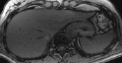
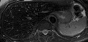
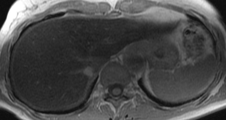
Marked signal loss of the liver parenchyma on in-phase T1-weighted GRE and T2 FSE images (use the spleen for comparison).
Case Discussion
There is a significant correlation between T1 and T2 relaxation rates and the liver iron content yielded by biopsy 1. Due to the absence of radiofrequency refocusing, gradient echo sequences are the most sensitive in detecting small amounts of iron 2. The signal changes here appear to be the opposite of those in hepatic steatosis. However, this phenomenon is not based on chemical shift, but on susceptibility effects, for the in-phase echo is the second echo of the double gradient-echo sequence, and is therefore acquired at a longer TE. This allows more time for dephasing due to field inhomogeneities.


 Unable to process the form. Check for errors and try again.