
Presentation
Abdominal pain
Patient Data
Age: 40 years
Gender: Female
From the case:
Hepatic alveolar echinococcosis


Show annotations
Download
Info
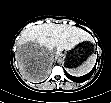
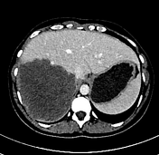
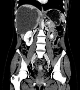
There is a large necrotic mass with irregular margins measuring 13.0 x 11.0 x 14.0 cm, with diffuse involvement of the right liver lobe. In the venous phase, there is no accumulation of contrast agent.
There are no calcifications and no extension into the hepatic portal vein.
Download
Info
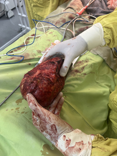
After surgery, histology confirms the diagnosis - hepatic alveolar echinococcosis.
Case Discussion
Hepatic alveolar echinococcosis is a very rare but aggressive and invasive form of hepatic hydatid disease caused by Echinococcus multilocularis. It presents with a wide variety of CT morphological manifestations. In this case, it shows infiltrative growth into the liver parenchyma.


 Unable to process the form. Check for errors and try again.