
Presentation
Right upper quadrant pain for few weeks.
Patient Data
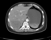

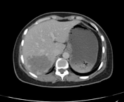
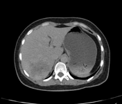

The liver is normally positioned and has normal size. A definable mass lesion of 6.0 x 7.0 x 6.3 cm (AP x TRANS x CC) is noted in segment VI of the liver which is showing, nodular and non-continuous, peripheral enhancement on arterial phase and progressive peripheral enhancement with more centripetal fill-in on venous and delayed phases, suggestive of hepatic hemangioma. At least three other lesions (larger one with similar characteristic) are noted in liver parenchyma as well.
A non-obstructive calculus of about 7.0 mm is noted at lower pole of the right kidney.
Evidence of pelvic congestion syndrome is appreciated.
Case Discussion
Characteristic imaging findings are in favor of hepatic haemangiomatosis along with right renal non-obstructive calculus and evidence of pelvic congestion syndrome as described above.


 Unable to process the form. Check for errors and try again.