
Presentation
Headache, fever and seizure of 1 week duration.
Patient Data


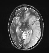
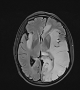
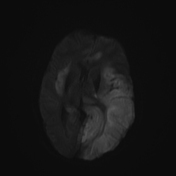
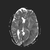
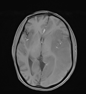
There are asymmetric T1 hypointense, T2/FLAIR hyperintense lesions involving the bilateral limbic system, insular cortex and part of the frontal lobe. Mass effect with effacement of the left lateral ventricle and diffusion restriction within the signal abnormality. The basal ganglia is spared. These findings are characteristic of herpes simplex encephalitis.
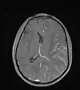
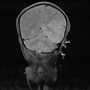
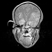
Post-contrast gyral and leptomeningeal enhancement, and extra-limbic involvement of the left parietal, and occipital lobe are also seen, which are less characteristic but also compatible with herpes simplex meningoencephalitis.
Case Discussion
Asymmetric T2/FLAIR hyperintense lesions involving the limbic system bilaterally, insular cortex and inferior part of the frontal lobes asymmetrically. The basal ganglia are spared, helping to exclude middle cerebral artery infarction. This pattern is characteristic of herpes simplex viral (HSV) encephalitis 1-4.
Mild post-contrast gyral and leptomeningeal enhancement suggest a meningitis component (meningoencephalitis) in this case 3,4. Extra-limbic involvement of the left parietal and occipital lobe are less characteristic but also compatible with a presentation of HSV encephalitis 1-3.
Although not demonstrated, hemorrhagic transformation is often seen in adult HSV encephalitis 1,4.


 Unable to process the form. Check for errors and try again.