
Presentation
Shortness of breath on exertion. Echocardiogram and brain natriuretic peptide (BNP) levels were normal. There was no history of smoking.
Patient Data
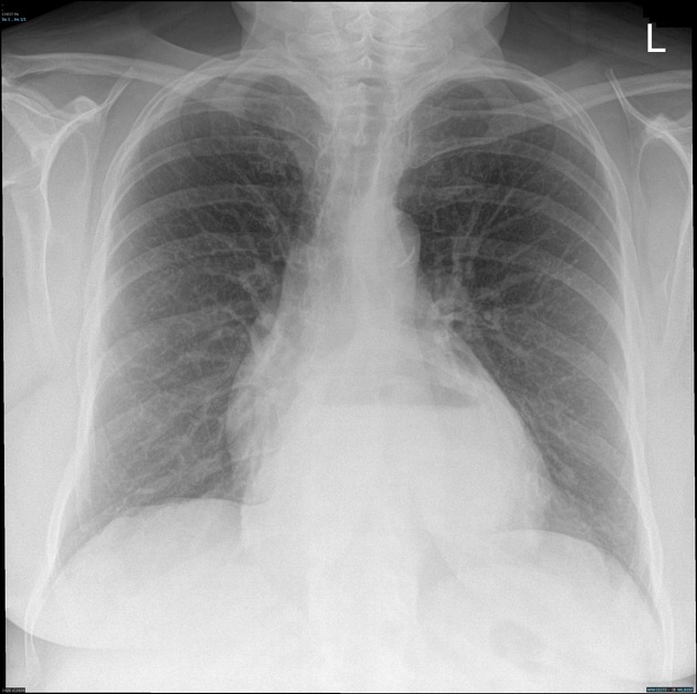
There is a hiatus hernia projected over the retrocardiac region. Air-fluid level is seen within the hiatus hernia. The heart and mediastinal contours are otherwise within normal limits. No focal active lung lesion or pleural effusion is demonstrated.
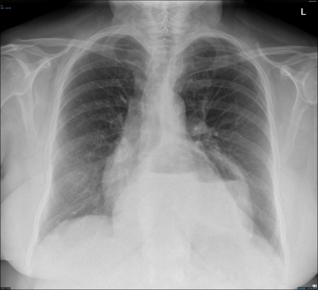
The hiatus hernia is larger as compared to the previous chest x-ray in year 2020. Mild cardiomegaly is noted.
Case Discussion
This is a case of hiatus hernia, which has increased in size after 4 years. The patient was referred to the upper gastrointestinal surgical clinic for surgical management.
Hiatus hernias happen when abdominal contents protrude through the esophageal opening of the diaphragm into the chest cavity.


 Unable to process the form. Check for errors and try again.