
Presentation
Routine bone density assessment. History of chronic kidney disease and hypercalcemia.
Patient Data
Dual-energy x-ray absorptiometry (DXA) of the lumbar spine and right hip.
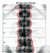
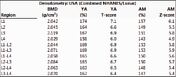
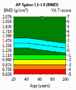
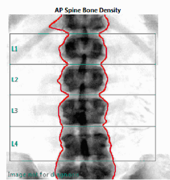
Anterior-posterior (AP) view of the lumbar spine demonstrates diffuse, homogeneously increased bone mineral density (BMD). There is no compression fracture(s). No overlying artifact. BMD of L1-4 measures 2.058 g/cm2 for a markedly elevated Z-score of 5.6.
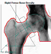
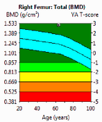
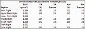
AP view of the right hip, acquired with mild internal rotation, shows upper limits of normal BMD with Z-scores of 2.0 for both the femoral neck and total femur regions of interest (ROIs).
Case Discussion
Unlike osteopenia and osteoporosis which are defined by the World Health Organization (WHO) using DXA T-scores, there is no WHO definition for high (supranormal) bone mineral density. Hence most authors classify it based upon the Z-score, which is based upon comparison to individuals of the same gender, age, race/ethnicity, and height/weight. A Z-score of >=2.5 is considered abnormal by many authors and termed high bone mineral density.
The differential for diffuse high bone mineral density is different from focal high bone mineral density, and also varies between adults and children. Given this patient's history of chronic kidney disease and hypercalcemia, this case is presumed to represent renal osteodystrophy.


 Unable to process the form. Check for errors and try again.