
Presentation
Headache and blurred vision. Hypothalamic tumour. Right frontal VAD. Biopsy. Left occipital VP shunt.
Patient Data


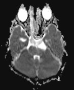
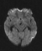
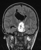
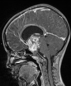
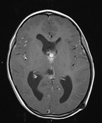
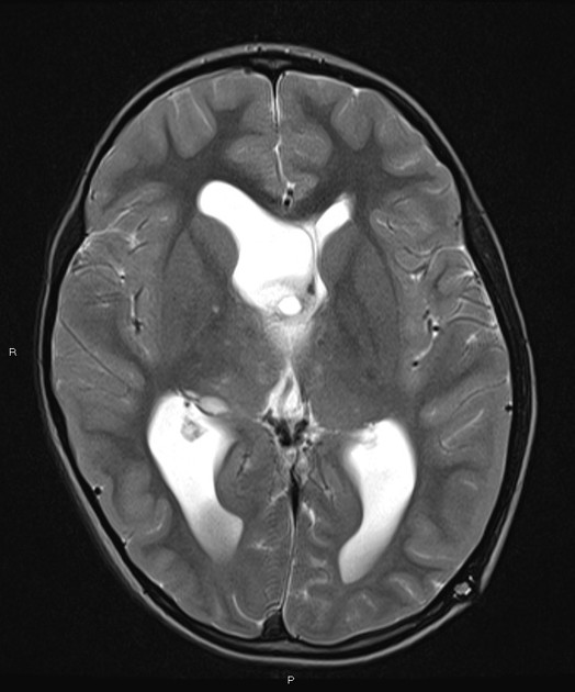
Large enhancing tumour centred in the hypothalamic region. Heterogeneous contrast enhancement. Obstructive hydrocephalus. No other abnormality.
Right frontal VAD. Left occipital VP shunt.
PATHOLOGY
Sections show fragments of a glial tumour composed of elongated bipolar piloid cells arranged in a composite solid and loose architecture. Nuclear pleomorphism is not marked.Mitotic activity is inconspicuous. No evidence of necrosis. No well-formed Rosenthal fibres or eosinophilic granular bodies.
Strong expression: glial fibrillary acidic protein and S100 protein
Negative reaction: antibodies to IDH1 and epithelial membrane antigen.
Cell proliferation index (MIB1 antibody) is overall low.
Pilocytic astrocytoma WHO Grade 1.


 Unable to process the form. Check for errors and try again.