
Presentation
Bilateral papilledema grade 3
Patient Data
Age: 20 years
Gender: Female
From the case:
Idiopathic intracranial hypertension


Download
Info
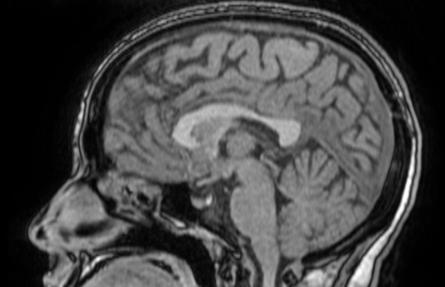
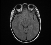
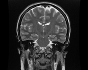
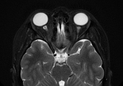
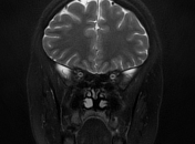
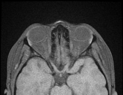
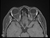
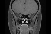
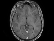
The MRI sequences demonstrate:
- flattening of the posterior sclera bilaterally
- protrusions of the optic nerve heads
- vertical tortuosity of the optic nerves
- enhancement of the prelaminar (intra-ocular) optic nerves well-visualized on postcontrast axial T1 fat saturation
- slight enlargement of the subarachnoid space around the optic nerves
- Partially empty sella turcica (pituitary gland's height=4.5 mm)
- the cerebral venous sinuses are patent on postcontrast sequences with no significant stenosis (MRV was not performed)
- no mass lesion is seen at the infra- or supratentorial level.
Case Discussion
MRI findings suggestive of an idiopathic intracranial hypertension
In the absence of a cause for intracranial hypertension, the imaging features should suggest an idiopathic origin


 Unable to process the form. Check for errors and try again.