
Presentation
Bilateral papilloedema grade 3
Patient Data
Age: 35 years
Gender: Female
From the case:
Idiopathic intracranial hypertension (IIH)


Download
Info
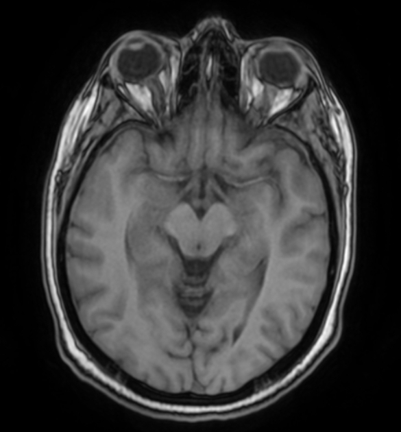
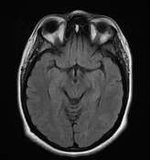
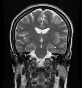
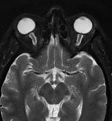
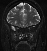
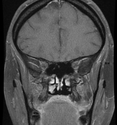
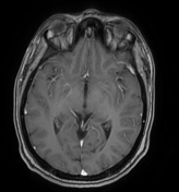
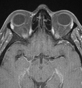
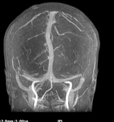
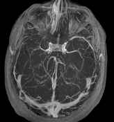
The MRI sequences demonstrate:
- vertical tortuosity of the optic nerves
- enlargement of the subarachnoid space around the optic nerves
- flattening of the posterior sclera bilaterally
- protrusions of the optic nerve head mainly on the right
- Partially empty sella turcica
- enlarged Meckel cave mainly on the left
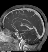
- the cerebral venous sinuses are patent on postcontrast sequences with stenosis in distal portions of both transverse sinuses on MRV images
- No mass lesion at infra-or supra-tentorial regions


 Unable to process the form. Check for errors and try again.