
Presentation
Right hip pain after fall.
Patient Data
Age: 60 years
Gender: Female
Download
Info
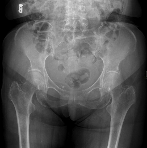
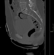
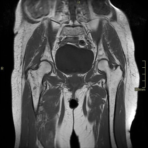
No fracture or dislocation.
From the case:
Intertrochanteric femur fracture - spectral CT


Show annotations
Download
Info

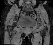
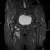
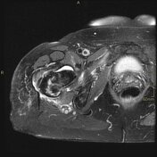
Bone marrow oedema in the right intertrochanteric region is seen on calcium suppression images.
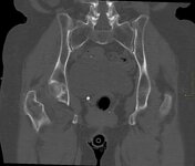
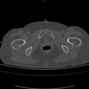
Subtle sclerosis in the intertrochanteric region is seen on coronal bone window images. No cortical breach was seen. Compare with the opposite site.
From the case:
Intertrochanteric femur fracture - spectral CT

Show annotations
Download
Info
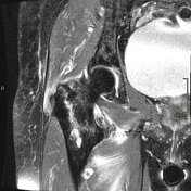
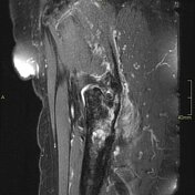
Intertrochanteric fracture line seen on T1 with fluid and oedema on STIR images.
Case Discussion
Calcium suppression images showing bone marrow oedema are useful in detecting occult nondisplaced acute fractures.
This is a useful application of spectral/dual energy CT.
MRI helped in confirming the diagnosis.


 Unable to process the form. Check for errors and try again.